
A diag image is a picture taken inside the body so that doctors can see what is happening. In fact, the term combines “diag” (short for diagnostic) and “image” (a visual picture). These pictures are made by special machines and help doctors find illness, injury or changes in the body that cannot be seen from the outside. In modern medicine, diag images are used routinely in hospitals and clinics to help doctors make smart decisions.
Why is a diag image important?
First, a diag image lets doctors look inside the body without surgery. Instead of cutting open someone to see bones, tissues or organs, the image shows what is going on. Because of that, less pain and risk are needed for patients. Also, diag images help in early detection of trouble, so treatment can begin sooner and outcomes can improve. Furthermore, these images allow doctors to monitor progress—if someone is sick and being treated, repeated diag images show whether the condition is improving or whether change is needed.
How is a diag image created?
Creating a diag image involves several steps. First, a doctor or technician decides what kind of image is needed—bones, brain, soft tissue, or organs. Then the patient is sent to the imaging room where a machine is used. Different machines work in different ways. For example:
An X-ray uses a small amount of radiation passing through the body to capture a flat image of bones and dense tissues.
A CT scan (computed tomography) uses many X-ray slices taken from different angles, then a computer combines them for a detailed 3-D-type view.
An MRI (magnetic resonance imaging) uses strong magnets and radio waves to image soft tissues like brain, muscles or joints. No X-ray radiation is used.
An ultrasound uses high-frequency sound waves to “see” inside without radiation; often used in pregnancy, organs and soft tissue.
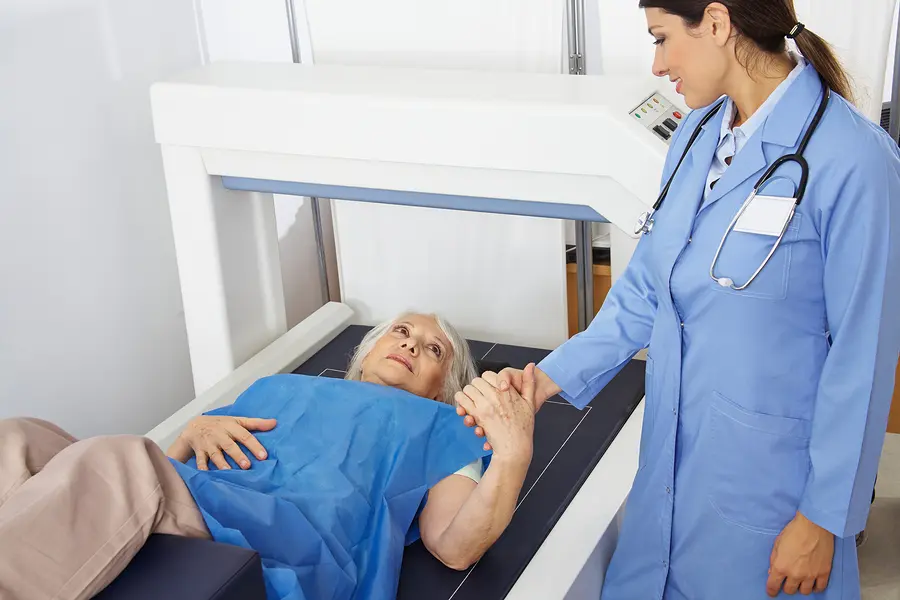
During the scan the patient may need to lie still, remove metal objects, or wear special clothing, depending on the technique. After the image is taken, the doctor or radiologist analyses it and writes a report. Then the treating doctor uses that report to decide what to do next.
What kinds of diag images exist?
There are many types of diag images. They differ by technology and by what part of the body they show. Some of the common types are:
X-ray images: good for viewing bones, fractures, chest organs.
CT scans: deeper view, useful for complex bones, internal organs, trauma.
MRI scans: best for soft-tissue detail such as brain, spine, joints.
Ultrasound: safe, portable, used for organs, pregnancies, and soft tissues.
PET scans and nuclear medicine: show how organs work rather than just how they look; useful in cancer, heart disease.
Each type has strengths and weaknesses. For example, X-rays are quick and cheap but less detailed for soft tissues. Meanwhile MRI is very detailed but takes longer and is more expensive.
How are diag images used in healthcare?
Diag images are used in many ways in medicine. First, they are used for diagnosis: when a patient has symptoms like pain, swelling or injury, a scan may reveal the cause. Then, they are used for treatment planning: before surgery or therapy, doctors check images to determine the best approach. After treatment, repeated imaging checks whether healing is working or whether a change is needed. Furthermore, in screenings (for example, breast cancer mammography) images are used to detect disease early before symptoms appear. Also, in emergencies, imaging may be used to quickly see internal injuries, bleeding or organ damage—so fast decisions can be made.
What are the benefits of diag images?
There are many benefits. Because the inside of the body can be seen, doctors can avoid exploratory surgery, thereby reducing risk and cost. Because problems are detected earlier, treatments can begin sooner, which often leads to better outcomes. Imaging also gives detailed information: for example, surgeons know exactly where to operate and how. Additionally, the patient experience is improved: many imaging tests are painless or minimally uncomfortable. Finally, imaging supports monitoring: doctors can track how treatment is working and adjust if needed. All of these benefits help make healthcare more precise, safe and effective.
What are the risks and limitations?
Although diag imaging is powerful, there are risks and limitations. Some imaging tests involve radiation (e.g., X-ray, CT) which means exposure to small amounts of ionizing radiation. Doctors try to minimize this. Also, some imaging tests are not suitable for all patients. For example, MRI machines use strong magnets so they may not be safe for people with certain implants. Some machines may make you uncomfortable (small space, noise, lying still for a long time). Also, an image may not show everything: sometimes findings are unclear, or a scan may not detect early disease, or may detect incidental findings that worry patients unnecessarily. Cost and availability are other issues: advanced imaging may be expensive and not available everywhere. In summary, imaging is a powerful tool but not perfect.
How should you prepare for a diag image?
Preparation depends on the type of scan. For many tests you may be asked to remove metal jewelry, wear a hospital gown, or refrain from eating for a few hours. For CT or MRI you might need to lie very still; for MRI you might be asked to remove all metal from your body because of the magnet. Contrast material (a dye) may need to be swallowed, injected, or introduced into your body to make certain structures visible. A technician or nurse will explain the process beforehand. Also you should tell your doctor if you are pregnant, have metal implants or have kidney problems (because of contrast material). Proper preparation makes the imaging safer, clearer and more accurate.
What do doctors look for in a diag image?
When a diag image is reviewed, doctors (or radiologists) look for abnormalities—areas that differ from what is normal for that part of the body. They examine shape, size, density, texture and movement (in some imaging like ultrasound). They compare with what is expected. For instance, a fracture shows a break in the bone line on an X-ray; a tumor may show as an unusual mass on MRI. Also they check for change over time: has something grown, shrunk, changed shape or responded to treatment? The findings are reported, often with suggestions for further action. The report will go to the doctor treating you who will decide what to do next based on the image plus all other tests.
How is technology changing diag images?
As technology advances, diag imaging is evolving quickly. Artificial intelligence (AI) and machine learning are being used to analyse images and detect patterns that may be hard to spot with the human eye. Digital imaging means high resolution, faster transfer of images and easier sharing among specialists. Also portable imaging devices are improving so scans can be done at point-of-care (for example, at the bedside or in remote settings). Furthermore, lower radiation techniques, better contrast materials and more patient-friendly machines (open MRI, faster scans) are being developed. Because of these trends, the power and accessibility of diag images are increasing, and they will likely play a bigger role in future healthcare.
What should a young person know about diag images?
If you are a teenager or soon will visit a clinic requiring a scan, here are some things to know:
Ask questions. If your doctor says “we’ll take a diag image”, ask why the image is needed and what the expected outcome is.
Be honest with your healthcare team. Let them know if you have metal in your body, if you are pregnant, or have any allergies (e.g., to contrast dye).
Stay still when instructed during the scan. Movement may blur the image and make repeat scanning necessary.
Understand that while diag images help, they are only one part of diagnosis. Your doctor will consider symptoms, medical history and other tests too.
If you feel anxious (some machines are loud or small), tell the technician. Many centres have options like music, ear plugs or even open scanners.
Support your health. A scan may show a problem, but strong lifestyle choices (good diet, exercise, sleep) matter for long-term wellness.
How much does a diag image cost and where is it done?
The cost and location vary widely by country, region, clinic and technology. Simple X-rays are usually low cost and widely available. More advanced scans (MRI, PET) cost more and may need referral to a specialist centre. In many countries public health insurance covers basic imaging; in others private payment or insurance is required. The procedure is usually done in a hospital, imaging centre or clinic. A referral from a doctor is often needed. Because of equipment and training costs, the availability of advanced imaging may be limited in rural or low-resource settings.
What is the future outlook for diag images?
Looking ahead, the role of diag images is expected to expand. With improved AI and deep learning, image interpretation may become faster and more accurate. Portable and low-cost imaging may reach remote and underserved areas, increasing access. Personalized imaging—tailored for individual patients based on their genetics, history and lifestyle—may become more common. Also, imaging may integrate more with other data (wearables, sensors, big data) so diagnosis and monitoring become more predictive rather than just reactive. Overall, diag images are likely to become even more vital in healthcare and our ability to manage health and disease will improve because of them.
Final-thought summary
In summary, a diag image is a powerful medical tool. It allows doctors to see inside the body, plan treatments, monitor progress and help patients get better results. The technology behind it is impressive and the applications are wide. At the same time, there are limitations—costs, availability, radiation risk and the need for proper interpretation. As a young person, knowing what diag images are and how they work helps you understand your health better and ask smart questions when they come up. Because health matters, the next time you hear the term diag image you’ll know that a lot of science, machines and care are behind that single scan.